Principles of Neuroimaging B - 2016: Difference between revisions
No edit summary |
|||
| (50 intermediate revisions by the same user not shown) | |||
| Line 1: | Line 1: | ||
Principles of Neuroimaging B, Winter, 2016 - Class Schedule and Syllabus | Principles of Neuroimaging B, Winter, 2016 - Class Schedule and Syllabus | ||
== This schedule ''will'' change!== | == This schedule ''will'' change!== | ||
| Line 44: | Line 43: | ||
=Week 2: MRI Techniques= | =Week 2: MRI Techniques= | ||
==''Monday 1/11/16'' == | ==''Monday 1/11/16'' == | ||
[[Image:ASL.png | right]] | |||
===- Measurement of Blood Flow by MRI. ''Danny JJ Wang'': [http://www.bri.ucla.edu/people/danny-jiong-jiong-wang-phd]=== | ===- Measurement of Blood Flow by MRI. ''Danny JJ Wang'': [http://www.bri.ucla.edu/people/danny-jiong-jiong-wang-phd]=== | ||
Most MRI images in use today are static maps, generally called "structural" images. However, the MRI signal is sensitive to a wide variety of dynamic phenomena. Practitioners typically use the term "functional" to describe images that pick up time-varying signals. The term ''functional MRI'', of course, has been usurped to describe brain activity mapping and, in most cases, refers to BOLD signal effects. | Most MRI images in use today are static maps, generally called "structural" images. However, the MRI signal is sensitive to a wide variety of dynamic phenomena. Practitioners typically use the term "functional" to describe images that pick up time-varying signals. The term ''functional MRI'', of course, has been usurped to describe brain activity mapping and, in most cases, refers to BOLD signal effects. | ||
Measurement of blood ''flow'' in particular is another window into brain activity. Several methods exist to create signal sensitive to blood flow, or blood flow changes. Some label the blood flow based on relaxation effects (Arterial Spin Labeling - ASL - being one important method) and others measure flow by velocity. | Measurement of blood ''flow'' in particular is another window into brain activity. Several methods exist to create signal sensitive to blood flow, or blood flow changes. Some label the blood flow based on relaxation effects (Arterial Spin Labeling - ASL - being one important method) and others measure flow by velocity. | ||
| Line 54: | Line 53: | ||
''Required Readings'' - Please complete these readings prior to class. | ''Required Readings'' - Please complete these readings prior to class. | ||
:*[[media:M284B_perfusion-web.pdf | Dr. Wang's Handout]] | :*[[media:M284B_perfusion-web.pdf | Dr. Wang's Handout]] | ||
<!-- | <!-- | ||
:*[http://www.cis.rit.edu/htbooks/mri/ These notes] by Joseph Hornak are highly professional and complete coverage of MRI.] | :*[http://www.cis.rit.edu/htbooks/mri/ These notes] by Joseph Hornak are highly professional and complete coverage of MRI.] | ||
| Line 63: | Line 59: | ||
''above'': Figure 1 from Hahn, 1950 | ''above'': Figure 1 from Hahn, 1950 | ||
--> | --> | ||
''Suggested Further Reading'' | ''Suggested Further Reading'' | ||
:*[http://www.pnas.org/content/89/1/212.long Williams, PNAS 1992] | :*[http://www.pnas.org/content/89/1/212.long Williams, PNAS 1992] | ||
| Line 69: | Line 64: | ||
:*[http://onlinelibrary.wiley.com/doi/10.1002/mrm.21790/epdf Dai, MRM 2008] | :*[http://onlinelibrary.wiley.com/doi/10.1002/mrm.21790/epdf Dai, MRM 2008] | ||
:*[http://www.ncbi.nlm.nih.gov/pubmed/17969096 Wu, MRM 2007] | :*[http://www.ncbi.nlm.nih.gov/pubmed/17969096 Wu, MRM 2007] | ||
:*[http://www.ncbi.nlm.nih.gov/pmc/articles/PMC3155947/ Zheng, Neuroimage 2012] | |||
==''Wednesday 1/13/16'' == | ==''Wednesday 1/13/16'' == | ||
| Line 102: | Line 98: | ||
:* | :* | ||
=Week 4: | =Week 4: Multimodal imaging= | ||
==''Monday 1/25/16'' == | ==''Monday 1/25/16'' == | ||
=== | ===Multimodal imaging. ''Speaker'': [http://www.brainmapping.org/MarkCohen MarkCohen]=== | ||
Handouts distributed in class. | Handouts distributed in class. | ||
''Required Readings'' - Please complete these readings prior to class. | ''Required Readings'' - Please complete these readings prior to class. | ||
:*[[media: | :*[[media:MultimodalNITP.pdf | slides handout]] | ||
<!-- | <!-- | ||
| Line 121: | Line 116: | ||
==''Wednesday 1/27/16'' == | ==''Wednesday 1/27/16'' == | ||
===Class canceled.=== | |||
=== | |||
=Week 5: | =Week 5: Positron Emission Tomography= | ||
[[Image:PET-ring.png | left]] | |||
==''Monday 2/1/16'' == | ==''Monday 2/1/16'' == | ||
=== | ===Positron Emission Tomography. ''Speaker'': [http://faculty.pharmacology.ucla.edu/institution/personnel?personnel_id=45558 Magnus Dahlboun]=== | ||
Handouts distributed in class. | |||
''Required Readings'' - Please complete these readings prior to class. | ''Required Readings'' - Please complete these readings prior to class. | ||
:* | :*[[media:PET_2016_M284B_SM.pdf | Dr. Dahlbom's PET handout]] | ||
<!-- | <!-- | ||
:*[http://www.cis.rit.edu/htbooks/mri/ These notes] by Joseph Hornak are highly professional and complete coverage of MRI.] | :*[http://www.cis.rit.edu/htbooks/mri/ These notes] by Joseph Hornak are highly professional and complete coverage of MRI.] | ||
| Line 142: | Line 136: | ||
:* | :* | ||
[[Image:PETfounders.png | right]] | |||
==''Wednesday 2/3/16'' == | ==''Wednesday 2/3/16'' == | ||
=== | ===PET applications. ''Speaker'': [http://www.semel.ucla.edu/profile/edythe-london Edythe London]=== | ||
''Required Readings'' - Please complete these readings prior to class. | ''Required Readings'' - Please complete these readings prior to class. | ||
:*[[Media:NITP_PET_2-3-16.pdf | Dr. Edythe London PET Slides]] <-- '''Modified 2-3-16''' | |||
:*[ | |||
-- | |||
''Suggested Further Reading'' | ''Suggested Further Reading'' | ||
=Week 6: | =Week 6: Brain Stimulation and Ultrasound= | ||
==''Monday 2/8/16'' == | ==''Monday 2/8/16'' == | ||
===- | ===TMS. ''Speaker'': [http://faculty.bri.ucla.edu/institution/personnel?personnel_id=121107 Allan Wu]=== | ||
'Required Readings'' | |||
:* [[media:NITP-talk-2016-02-08-distribution_SM.pdf | Wu Lecture slides 2016]] '''<- modified 2-9-2016''' | |||
:* [[media:TMSSafetyAndEthics-Rossi.pdf | TMS Safety and Ethics - Rossi, 2009]] | |||
:* [http://www.sciencedirect.com/science?_ob=ArticleURL&_udi=B6T0J-50CV801-1&_user=4423&_coverDate=01%2F31%2F2011&_rdoc=1&_fmt=high&_orig=search&_origin=search&_sort=d&_docanchor=&view=c&_acct=C000059605&_version=1&_urlVersion=0&_userid=4423&md5=f9fa3c7d63942dfb3c74e047a1f848bc&searchtype=a | M Sandrini, C Umilta and E Rusconi, “''The use of transcranial magnetic stimulation in cognitive neuroscience: a new synthesis of methodological issues.''” '''Neurosci Biobehav Rev''', '''35'''(3): p. 516-536. 2011] | |||
''Suggested Further Reading'' | ''Suggested Further Reading'' | ||
:* | Once upon a time we demonstrated that this sort of magnetic stimulation can take place in the MRI machines: | ||
:*[http://onlinelibrary.wiley.com/doi/10.1002/mrm.1910140226/pdf MS Cohen, RM Weisskoff, RR Rzedzian and HL Kantor, “Sensory stimulation by time-varying magnetic fields.” Magnetic Resonance in Medicine, 14(2): p. 409-414. 1990] | |||
==''Wednesday 2/10/16'' == | ==''Wednesday 2/10/16'' == | ||
=== | [[Image:Cetacean.png | right]] | ||
===Ultrasound. ''Speaker'': [https://www.linkedin.com/pub/george-saddik/2b/558/49 George Saddik]=== | |||
''Required Readings'' | |||
:*[[media:US_Imaging_Systems_1.pdf | Ultrasound Slides (2016)]] | |||
''Suggested Further Reading'' | ''Suggested Further Reading'' | ||
:* | :* [[media:Culjat2010.pdf | A review of tissue substitutes for ultrasound imaging ]] | ||
:*[[media:SaddikUltrasound2015.pdf | Ultrasound Slides (2015)]] | |||
=Week 7: | =Week 7: Optical neuroimaging and optogenetics= | ||
==''Monday 2/15/16'' == | ==''Monday 2/15/16'' == | ||
===- President's Day - School holiday: === | ===- President's Day - School holiday: === | ||
==''Wednesday 2/17/16'' == | ==''Wednesday 2/17/16'' == | ||
===- Optical Neuroimaging and Optogenetics. ''Peyman Golshani | [[Image:PTEN.png | left]] | ||
===- Optical Neuroimaging and Optogenetics. ''Speaker'': [http://golshanilab.neurology.ucla.edu Peyman Golshani]: === | |||
''Required Readings'' - Please complete these readings prior to class. | ''Required Readings'' - Please complete these readings prior to class. | ||
:* | :*[[media:Golshani_NITP_2016.pdf | Dr. Golshani's Methods slides]] | ||
:*[[media:PeymanResearchTalk.pdf | Dr. Golshani's Research slides]] | |||
:*[[media: | |||
''Suggested Further Reading'' | ''Suggested Further Reading'' | ||
:* | :* | ||
| Line 206: | Line 183: | ||
=Week 8: Advanced MRI methods= | =Week 8: Advanced MRI methods= | ||
==''Monday 2/22/16'' == | ==''Monday 2/22/16'' == | ||
===- Diffusion | [[Image:DIffusingSpheres.png | left]] | ||
===- Diffusion Physics ''Speaker'': [http://radiology.ucla.edu/body.cfm?id=48&action=detail&ref=18 Ben Ellingson]=== | |||
''Required Readings'' | |||
Slides should be up soon. | |||
:*[https://www.dropbox.com/s/0feqz7tlojbfgzl/1_DiffusionMRI_Methods_2015.key.pdf?dl=0 Handouts for class on 1/14/15] ''<- New 1/26-2015'' | |||
:*[[media:1_DiffusionMRI_Methods_v1 - 2014.pdf | Ben Ellingson Diffusion Slides]] uploaded 3/14/14 | |||
''Suggested Further Reading'' | |||
:*[[media:DTIHageman.pdf | Hageman DTI Slides]] | |||
:*[[media:NICourse_DTILecture4Post_11-01-26.pdf | Hageman DTI Notes 2/26/12]] | |||
:*[http://www.ncbi.nlm.nih.gov/pubmed/15037456?dopt=Citation Diffusion tensor imaging of cerebral white matter: a pictorial review of physics, fiber tract anatomy, and tumor imaging patterns AJNR Am J Neuroradiol. 2004 Mar;25(3):356-69] | |||
:Sadly, the library does not have a subscription for the journals below (Mark has copies on reserve in his office): | |||
: | :*[http://www.ncbi.nlm.nih.gov/pubmed/8661285?dopt=Citation Microstructural and physiological features of tissues elucidated by quantitative-diffusion-tensor MRI. J Magn Reson B. 1996 Jun;111(3):209-19] | ||
:*[http://www.ncbi.nlm.nih.gov/pubmed/16950152?dopt=Citation Principles of diffusion tensor imaging and its applications to basic neuroscience research. Neuron. 2006 Sep 7;51(5):527-39] | |||
:*[http://www. | |||
:*[ | |||
- | |||
==''Wednesday 2/25/16'' == | ==''Wednesday 2/25/16'' == | ||
===- Brain Morphometry. ''Roger Woods | [[Image:Callosum.png | right]] | ||
===- Brain Morphometry. ''Speaker'': [http://www.bmap.ucla.edu/about/peopledetails/roger_woods/ Roger Woods]: === | |||
''Required Readings'' - Please complete these readings prior to class. | |||
:*[[media:M284B_Woods.pdf | Dr. Woods' slides]] | |||
''Suggested Further Reading'' | ''Suggested Further Reading'' | ||
:* | :[http://www.ncbi.nlm.nih.gov/pubmed/12667854 *RP Woods, “Characterizing volume and surface deformations in an atlas framework: theory, applications, and implementation.” Neuroimage, 18(3): p. 769-788. 2003] | ||
=Week 9: MR Spectroscopy= | =Week 9: MR Spectroscopy= | ||
==''Monday 2/29/16'' == | ==''Monday 2/29/16'' == | ||
=== | No class | ||
==''Wednesday 3/2/16'' == | |||
=== Spectroscopy fundamentals. ''Speaker'': [http://people.healthsciences.ucla.edu/institution/personnel?personnel_id=74852 Joseph O'Neill] === | |||
''Required Readings'' - Please complete these readings prior to class. | ''Required Readings'' - Please complete these readings prior to class. | ||
:*[[media:ONeill2016Spectro.pdf | Dr. O'Neill's slides]] | |||
:*[[media: | |||
===- Spectroscopic Methods. ''Speaker'': [http://radiology.ucla.edu/body.cfm?id=48&action=detail&ref=65 Albert Thomas] === | |||
===- Spectroscopic Methods. '' | |||
''Required Readings'' - Please complete these readings prior to class. | ''Required Readings'' - Please complete these readings prior to class. | ||
:*[[media:M284B_AThomas030216.pdf | Dr. Thomas' slides]] | |||
:*[[media: | |||
''Suggested Further Reading'' | ''Suggested Further Reading'' | ||
:* | :* [http://www.ncbi.nlm.nih.gov/pubmed/23292856 SA Wijtenburg, LM Rowland, RA Edden and PB Barker, “Reproducibility of brain spectroscopy at 7T using conventional localization and spectral editing techniques.” J Magn Reson Imaging, 38(2): p. 460-467. 2013. PMCID: PMC3620961] | ||
:* [http://www.ncbi.nlm.nih.gov/pubmed/20574964 S Lipnick, G Verma, S Ramadan, J Furuyama and MA Thomas, “Echo planar correlated spectroscopic imaging: implementation and pilot evaluation in human calf in vivo.” Magn Reson Med, 64(4): p. 947-956. 2010. PMCID: PMC2946523] | |||
:* [http://www.ncbi.nlm.nih.gov/pubmed/22505247 JK Furuyama, NE Wilson, BL Burns, R Nagarajan, DJ Margolis and MA Thomas, “Application of compressed sensing to multidimensional spectroscopic imaging in human prostate.” Magn Reson Med, 67(6): p. 1499-1505. 2012.] | |||
=Week 10: Multimodal Methods= | =Week 10: Multimodal Methods / Course wrap up= | ||
==''Monday 3/7/16'' == | ==''Monday 3/7/16'' == | ||
===- Imaging Genetics. ''Carrie Bearden | [[Image:Kinship.png | right]] | ||
===- Imaging Genetics. ''Speaker'': [http://www.semel.ucla.edu/bearden-lab Carrie Bearden]: === | |||
''Required Readings'' - Please complete these readings prior to class. | |||
:*[[media:Bearden_NITP2_2016-3-4.pdf | Dr. Bearden's slides]] | |||
''Suggested Further Reading'' | ''Suggested Further Reading'' | ||
:* | :*[[media:Carter-Genomics.pdf | ''Enhancing the Informativeness and Replicability of Imaging Genomics Studies'' Carter et al.]] | ||
:*[[media:Flint_and_Geschwind.pdf | ''Genetics and genomics of psychiatric disease'' Flint and Geschwind]] | |||
==''Wednesday 3/9/16'' == | ==''Wednesday 3/9/16'' == | ||
===- | ===- Course wrap up and review. ''Mark Cohen'': [http://www.brainmapping.org/MarkCohen Mark Cohen]=== | ||
Latest revision as of 00:08, 10 March 2016
Principles of Neuroimaging B, Winter, 2016 - Class Schedule and Syllabus
This schedule will change!
Lecture Videos
- Hopefully, we can start recording the class sessions for those who cannot make a few lectures.
Course Reading
Required Reading
- Signal Processing for Neuroscientists by Wim van Drongelen
- This can be found as a PDF on scribd.com, for a small fee of $8.99
Supplemental Reading
- Matlab for Neuroscientists
- Link for download found here for a small fee: http://www.scribd.com/doc/88212458/Matlab-Matlab-for-Neuroscientists
- Cartoon Guide to Statistics
- Link for download found here for a small fee: http://www.scribd.com/doc/148072668/Cartoon-Guide-to-Statistics
- NOTE: if you subscribe for a Scribd account for a day, you can download as many documents as you like for one fee.
Week 1: Magnetic Resonance Imaging
Monday 1/4/16
- MRI Contrast. Mark Cohen: Mark Cohen
Magnetic Resonance Imaging (MRI) is probably the most influential and most flexible current means of imaging the human brain. It features a vast number of separable contrast mechanisms, and a near ideal combination of non-invasiveness, safety, resolution and metric accuracy. However, it is extraordinarily expensive and has limited temporal resolution, especially for functional studies
Required Readings
- MRI Slides
- These notes by Joseph Hornak are highly professional and complete coverage of MRI.
Suggested Further Reading
- eMRI is another excellent online MRI learning resource
- Erwin Hahn - Spin Echoes: Essential reading for the MRI community
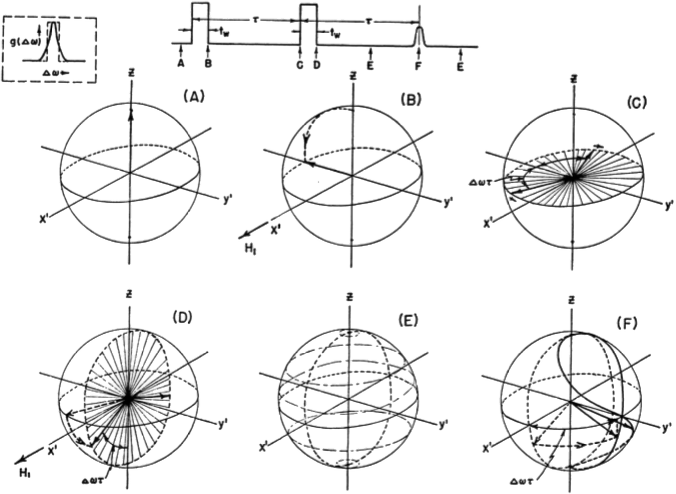
}
Wednesday 1/6/16
- MRI Image Formation and fMRI. Mark Cohen: Mark Cohen
Week 2: MRI Techniques
Monday 1/11/16
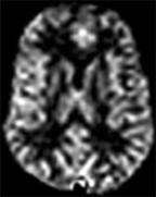
- Measurement of Blood Flow by MRI. Danny JJ Wang: [1]
Most MRI images in use today are static maps, generally called "structural" images. However, the MRI signal is sensitive to a wide variety of dynamic phenomena. Practitioners typically use the term "functional" to describe images that pick up time-varying signals. The term functional MRI, of course, has been usurped to describe brain activity mapping and, in most cases, refers to BOLD signal effects.
Measurement of blood flow in particular is another window into brain activity. Several methods exist to create signal sensitive to blood flow, or blood flow changes. Some label the blood flow based on relaxation effects (Arterial Spin Labeling - ASL - being one important method) and others measure flow by velocity.
In most cases, MRI is used as a semi quantitative modality. We get accurate spatial metrics but, in general, the image intensities are in arbitrary units. Blood flow is one particular feature that we can measure in native units.
Required Readings - Please complete these readings prior to class.
Suggested Further Reading
Wednesday 1/13/16
- More MRI. Mark Cohen: Mark Cohen
Required Readings - Please complete these readings prior to class.
Suggested Further Reading
Week 3: tbd
Monday 1/18/16
- MLK School Holiday
Wednesday 1/20/16
- tbd. Speaker: Mark Cohen
Abstract
Required Readings - Please complete these readings prior to class.
Suggested Further Reading
Week 4: Multimodal imaging
Monday 1/25/16
Multimodal imaging. Speaker: MarkCohen
Handouts distributed in class.
Required Readings - Please complete these readings prior to class.
Suggested Further Reading
Wednesday 1/27/16
Class canceled.
Week 5: Positron Emission Tomography
Monday 2/1/16
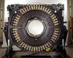
Positron Emission Tomography. Speaker: Magnus Dahlboun
Handouts distributed in class.
Required Readings - Please complete these readings prior to class.
Suggested Further Reading
Wednesday 2/3/16
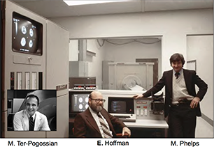
PET applications. Speaker: Edythe London
Required Readings - Please complete these readings prior to class.
- Dr. Edythe London PET Slides <-- Modified 2-3-16
Suggested Further Reading
Week 6: Brain Stimulation and Ultrasound
Monday 2/8/16
TMS. Speaker: Allan Wu
'Required Readings
Suggested Further Reading Once upon a time we demonstrated that this sort of magnetic stimulation can take place in the MRI machines:
Wednesday 2/10/16
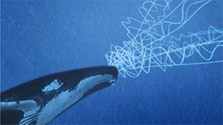
Ultrasound. Speaker: George Saddik
Required Readings
Suggested Further Reading
Week 7: Optical neuroimaging and optogenetics
Monday 2/15/16
- President's Day - School holiday:
Wednesday 2/17/16
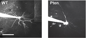
- Optical Neuroimaging and Optogenetics. Speaker: Peyman Golshani:
Required Readings - Please complete these readings prior to class.
Suggested Further Reading
Week 8: Advanced MRI methods
Monday 2/22/16
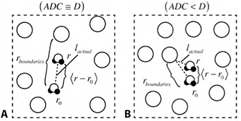
- Diffusion Physics Speaker: Ben Ellingson
Required Readings Slides should be up soon.
- Handouts for class on 1/14/15 <- New 1/26-2015
- Ben Ellingson Diffusion Slides uploaded 3/14/14
Suggested Further Reading
- Sadly, the library does not have a subscription for the journals below (Mark has copies on reserve in his office):
Wednesday 2/25/16
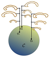
- Brain Morphometry. Speaker: Roger Woods:
Required Readings - Please complete these readings prior to class.
Suggested Further Reading
Week 9: MR Spectroscopy
Monday 2/29/16
No class
Wednesday 3/2/16
Spectroscopy fundamentals. Speaker: Joseph O'Neill
Required Readings - Please complete these readings prior to class.
- Spectroscopic Methods. Speaker: Albert Thomas
Required Readings - Please complete these readings prior to class.
Suggested Further Reading
- SA Wijtenburg, LM Rowland, RA Edden and PB Barker, “Reproducibility of brain spectroscopy at 7T using conventional localization and spectral editing techniques.” J Magn Reson Imaging, 38(2): p. 460-467. 2013. PMCID: PMC3620961
- S Lipnick, G Verma, S Ramadan, J Furuyama and MA Thomas, “Echo planar correlated spectroscopic imaging: implementation and pilot evaluation in human calf in vivo.” Magn Reson Med, 64(4): p. 947-956. 2010. PMCID: PMC2946523
- JK Furuyama, NE Wilson, BL Burns, R Nagarajan, DJ Margolis and MA Thomas, “Application of compressed sensing to multidimensional spectroscopic imaging in human prostate.” Magn Reson Med, 67(6): p. 1499-1505. 2012.
Week 10: Multimodal Methods / Course wrap up
Monday 3/7/16
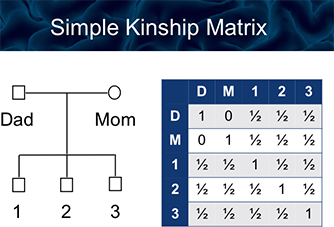
- Imaging Genetics. Speaker: Carrie Bearden:
Required Readings - Please complete these readings prior to class.
Suggested Further Reading